What Is Cervical Foraminal Stenosis — and Why the Differential Diagnosis Matters

The foramen is the bony channel through which each cervical nerve root exits the spinal canal. Cervical foraminal stenosis is the narrowing of that channel from degenerative changes — specifically, bone spur formation from uncovertebral joint hypertrophy and facet joint arthritis, which progressively encroach on the nerve root's exit space. Unlike disc herniation, which is often acute in onset, foraminal stenosis from bone tends to develop gradually and produces symptoms that worsen slowly over months to years. The clinical presentation is cervical radiculopathy — pain, numbness, tingling, and weakness in the arm and hand in the distribution of the affected nerve root. What distinguishes foraminal stenosis from disc herniation clinically is the chronicity: stenosis-driven symptoms tend to be more gradual in onset, more persistent rather than episodic, and less likely to resolve spontaneously than acute disc herniation.

The symptoms also tend to be more consistently present with sustained positions — arms overhead, prolonged typing, extended driving — rather than acute and severe as with large disc herniations. The differential diagnosis is genuinely important here. Carpal tunnel syndrome compresses the median nerve at the wrist and produces numbness and tingling in the thumb, index, and middle fingers — overlapping significantly with C6 radiculopathy. Thoracic outlet syndrome can produce diffuse arm symptoms. Ulnar nerve entrapment at the elbow overlaps with C8 radiculopathy. Shoulder pathology can produce referred arm pain. Getting to the right answer requires systematic clinical evaluation and, in ambiguous cases, electrodiagnostic testing.

How Integrated Care Improves the Differential Diagnosis Process

For patients within the Inspira Health Network who have been evaluated for arm and hand symptoms, the journey to our Mullica Hill spine office often includes documentation of prior workup: nerve conduction studies ordered by an Inspira neurologist, shoulder imaging from an Inspira orthopedist, or primary care notes documenting the evolution of symptoms over time. That prior evaluation — rather than representing redundant testing — often gives us important differential information before we even begin our examination.

Our evaluation begins with a systematic neurological examination: testing each cervical nerve root level's sensory distribution, motor strength, and reflex pattern. We assess Spurling's sign (neck extension and lateral bending that reproduces arm symptoms suggests cervical origin), ulnar versus median versus radial sensory territories, and the shoulder examination to exclude rotator cuff and acromioclavicular joint pathology as contributors. When the clinical picture is not definitive, electrodiagnostic testing — EMG and nerve conduction velocity — helps localize the lesion to the nerve root level rather than a peripheral site.

MRI of the cervical spine with attention to foraminal dimensions at each level provides the structural correlate for the clinical findings. When imaging and examination point to the same nerve level, the diagnostic picture is complete. When they diverge — when imaging shows multiple levels of foraminal narrowing but the examination suggests only one is clinically active — the electrodiagnostic study is the tiebreaker.

Treatment for Cervical Foraminal Stenosis: From Conservative to Surgical

Conservative management is the appropriate first approach for most patients. Because foraminal stenosis from bone spur formation does not spontaneously resolve the way disc herniation can, the goal of conservative management is different: symptom control and functional maintenance rather than structural resolution. Physical therapy focused on cervical positioning, postural mechanics, and nerve mobility exercises can meaningfully improve symptoms. Cervical epidural steroid injections, targeted at the affected foraminal level, can provide useful symptom relief in appropriate patients. When conservative measures are insufficient — when symptoms are progressive, when neurological deficits are advancing, or when the patient's function is significantly impaired — surgical decompression is appropriate. Posterior cervical foraminotomy, which widens the narrowed foramen through a small posterior incision without fusion, is the procedure most specifically suited to foraminal stenosis when disc height is preserved and there is no instability. ACDF is appropriate when disc space collapse, instability, or the anatomical pattern of compression make an anterior approach more appropriate.

Background media

Your Surgical Team at POA Mullica Hill

Rahul V. Shah

Dr. Rahul V. Shah, MD, FAAOS

Board-Certified Orthopaedic Spine Surgeon  |  Fellowship-Trained, UCSF  |  Chief Resident, Yale

Dr. Shah's approach to cervical foraminal stenosis is built on precise clinical correlation. His training at UCSF emphasized that imaging findings guide but do not dictate — a foraminal that looks narrow on MRI is only clinically significant if the nerve at that level is actually symptomatic. He applies a systematic examination to identify the level where the clinical signal is loudest, uses electrodiagnostics when the picture is ambiguous, and makes surgical recommendations based on the convergence of clinical, electrophysiological, and imaging evidence. For Mullica Hill patients who have been through the Inspira neurology or PM&R pathway before reaching our office, his evaluation builds on that prior workup rather than starting from scratch.

  • FAAOS — Fellow, American Academy of Orthopaedic Surgeons
  • Spine Surgery Fellowship — University of California, San Francisco
  • Chief Resident — Yale University Orthopaedic Surgery
  • Faculty — Rowan University Medical School

View Full Profile

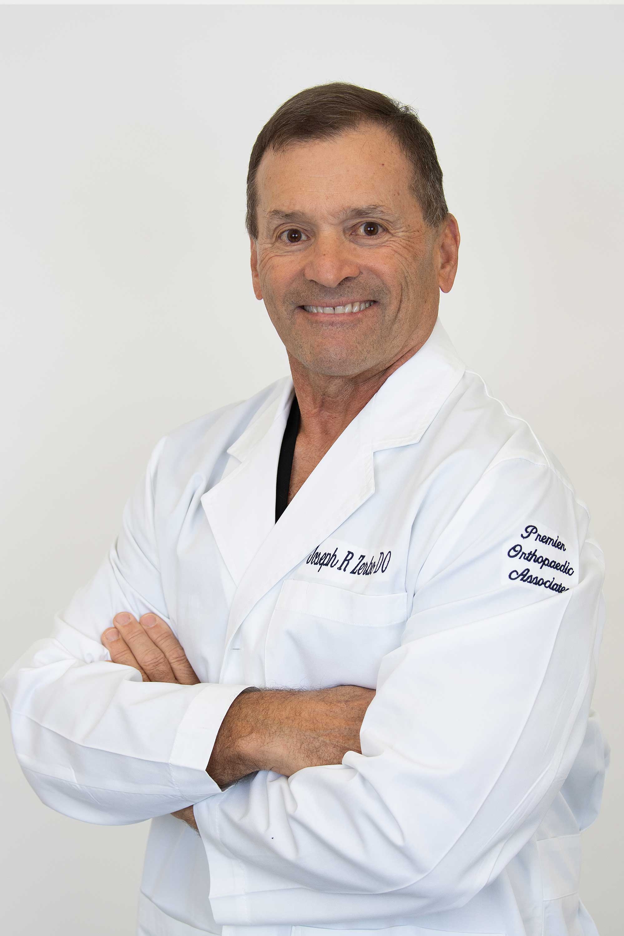
Joseph R. Zerbo male in white lab coat

Dr. Joseph R. Zerbo, DO

Board-Certified Orthopaedic Spine Surgeon  |  NASS Member  |  35+ Years in Southern NJ

Dr. Zerbo values the integrated care advantage that the Inspira network provides for cervical foraminal stenosis patients in particular — because the differential diagnosis here benefits from multi-specialty evaluation, and when neurology, primary care, and orthopedic spine surgery are all operating within the same system, the communication that resolves ambiguous cases happens more efficiently. His D.O. whole-spine orientation also means he examines the cervical spine in the context of the entire axial skeleton: thoracic kyphosis, shoulder mechanics, and overall posture all influence how cervical nerve roots are loaded, and his assessment reflects that broader anatomical picture.

  • AOBOS Board-Certified — American Osteopathic Board of Orthopaedic Surgeons
  • NASS Member — North American Spine Society (since 1999)
  • Chief Resident — Kennedy Memorial Hospital / UMDNJ
  • Founder — Zerbo Spine, PA

View Full Profile

ALIF FAQs

I was evaluated for carpal tunnel syndrome but the treatment didn't help — could this be my cervical spine?

Can cervical foraminal stenosis get better on its own?

What does electrodiagnostic testing add to an MRI in evaluating cervical foraminal stenosis?

Is foraminotomy the right surgery for cervical foraminal stenosis?

I was evaluated for carpal tunnel syndrome but the treatment didn't help — could this be my cervical spine?

Yes, and this is a common presentation. Carpal tunnel syndrome and C6 cervical radiculopathy produce very similar symptom patterns — numbness and tingling in the thumb and index finger, weakness in grip. If carpal tunnel treatment (splinting, injection, or surgery) did not resolve your symptoms, cervical foraminal stenosis at C6 is a diagnosis worth evaluating. A structured cervical neurological examination and MRI, combined with nerve conduction studies, can distinguish between the two. Some patients have both conditions simultaneously — a double crush phenomenon — which is why treatment of one does not always fully resolve the symptoms.

Can cervical foraminal stenosis get better on its own?

Disc herniations causing radiculopathy often improve spontaneously as the disc material resorbs over weeks to months. Foraminal stenosis from bone spur formation does not resorb — the structural narrowing is a fixed bony change. What can improve is the level of nerve root inflammation and sensitization, which means symptoms may fluctuate. Patients typically find that positioning, activity modification, and conservative treatment keep symptoms manageable for periods. However, bone-driven foraminal stenosis tends to be progressive over a long enough timeline, and if neurological deficits are advancing, earlier intervention is appropriate.

What does electrodiagnostic testing add to an MRI in evaluating cervical foraminal stenosis?

MRI shows the anatomy — where the foramen is narrow and where the nerve root appears compressed. Electrodiagnostic testing (EMG and nerve conduction velocity) shows function — whether the nerve at a given level is actually conducting normally or shows evidence of injury. This distinction matters when imaging shows multiple levels of narrowing, because surgery should be directed at the clinically active level, not every level that appears narrowed on imaging. Electrodiagnostics help localize the primary source of nerve dysfunction when imaging alone cannot resolve it.

Is foraminotomy the right surgery for cervical foraminal stenosis?

It is the most specifically targeted surgery for foraminal stenosis when the anatomy supports it. Foraminotomy directly widens the narrowed exit channel from a posterior approach without fusion — which means it addresses the structural problem precisely while preserving motion at the treated level. The criteria for foraminotomy candidacy are: predominantly foraminal compression rather than central disc herniation, preserved disc height, and no segmental instability. When those criteria are met, foraminotomy is an excellent procedure for cervical foraminal stenosis. When they are not, ACDF may be more appropriate — and we explain the anatomical reasoning for whichever approach we recommend.
Premier Orthopaedic Associates Mullica Hill - Inspira Hospital

Contact

(856) 690-1616

Hours of Operation

MON — FRI: 8:30AM - 5PM

Contact This Location
Contact us media
Accessibility: If you are vision-impaired or have some other impairment covered by the Americans with Disabilities Act or a similar law, and you wish to discuss potential accommodations related to using this website, please contact our Accessibility Manager at (856) 690-1616.
How can I help you?

Hey there, how can I help you?

Schedule a Visit Find a Provider Bill Pay
Contact Us