Lumbar Disc Herniation and Sciatica: The Problem Microdiscectomy Solves

The lumbar discs — the cartilaginous cushions between the vertebrae in the lower back — are subject to decades of compressive loading and gradual degeneration. When the outer fibrous layer of a disc (the annulus fibrosus) tears, the softer inner nucleus can protrude through the tear. If that protrusion contacts a nerve root exiting the spinal canal, the result is radiculopathy: the sharp, electric, often debilitating pain that follows the path of the compressed nerve from the low back through the buttock and down the leg into the foot. This is sciatica. The intensity of sciatica is frequently disproportionate to how it sounds to someone who has never experienced it.

Patients describe being unable to sit, unable to sleep, unable to find any position that provides relief. Work becomes impossible. The quality of life impact of severe lumbar radiculopathy is as significant as many chronic medical conditions, and for patients in whom conservative care has failed, it is not a condition to simply endure. Microdiscectomy removes the fragment of disc that is pressing on the nerve root. It does not remove the entire disc. It targets precisely what is causing the nerve compression and removes only that — leaving the disc itself and the structural integrity of the segment as intact as possible.

When Is Microdiscectomy the Right Treatment?

Microdiscectomy is recommended at our Cherry Hill office when the following conditions are met:

  • A herniated lumbar disc confirmed on MRI corresponds to the patient's specific pattern of leg pain and neurological symptoms
  • An adequate conservative trial — typically six to twelve weeks of physical therapy and/or epidural steroid injection — has not produced sufficient relief
  • Symptoms are causing a meaningful functional limitation (inability to work, loss of sleep, severe activity restriction)
  • No contraindication to surgery exists based on the patient's medical history
  • The timeline shortens when progressive neurological deficits are present — weakness in the leg or foot, loss of bowel or bladder function. These findings create urgency. Cauda equina syndrome — bilateral leg weakness with bowel and bladder involvement — is a surgical emergency that requires immediate decompression rather than a trial of conservative care.

Microdiscectomy is not recommended for back pain alone, for disc herniations found incidentally on imaging without corresponding symptoms, or for patients who have not yet had an appropriate conservative trial except in urgent circumstances. At our Cherry Hill office, the threshold is applied consistently: the right operation at the right time for the right patient.

Background media

What Happens During a Microdiscectomy?

Microdiscectomy is performed under general anesthesia with the patient prone. A small incision — typically 1 to 1.5 inches — is made in the lower back over the affected level. Using surgical loupes or a microscope for magnification, the surgeon gently moves the paraspinal muscles aside, makes a small opening in the ligamentum flavum, and directly visualizes the herniated disc fragment and the compressed nerve root.

The nerve root is carefully retracted, and the herniated fragment is removed with precision instruments. Any additional fragments within the disc space that could re-herniate are assessed and removed as appropriate. The decompressed nerve root is then confirmed to be free of any residual compression. The wound is closed in layers.

The entire procedure typically takes 45 minutes to one hour. Most Cherry Hill patients undergo microdiscectomy at Millennium Surgical Center as an outpatient and go home the same day.

Microdiscectomy Recovery: What to Expect

Microdiscectomy recovery is one of the most rewarding aspects of this procedure — the improvement in sciatica symptoms is often immediate and dramatic. Most patients report that the leg pain that brought them to surgery is significantly reduced or gone within the first 24 to 48 hours. The back soreness from the incision itself resolves over one to two weeks.

  • Days 1–7: Walking encouraged from day one. Light activity, wound care, gradual mobility. Most patients are off narcotic pain medication within a few days.
  • Weeks 1–3: Return to driving once off narcotics and cleared. Return to desk work typically in this window.
  • Weeks 3–6: Physical therapy begins, focusing on lumbar stabilization, flexibility, and gradual return to loading.
  • Weeks 6–12: Return to full activity including exercise, labor-intensive work, and sport for most patients.

The risk of re-herniation at the same level is approximately 5–10% over the lifetime of the disc. Patients who maintain core strength, manage body weight, and follow activity guidelines have meaningfully lower re-herniation rates. For Cherry Hill patients who re-herniate after an initial successful microdiscectomy, revision microdiscectomy or fusion at that level may be discussed depending on the circumstances.

Banner media

Your Surgical Team at POA Cherry Hill

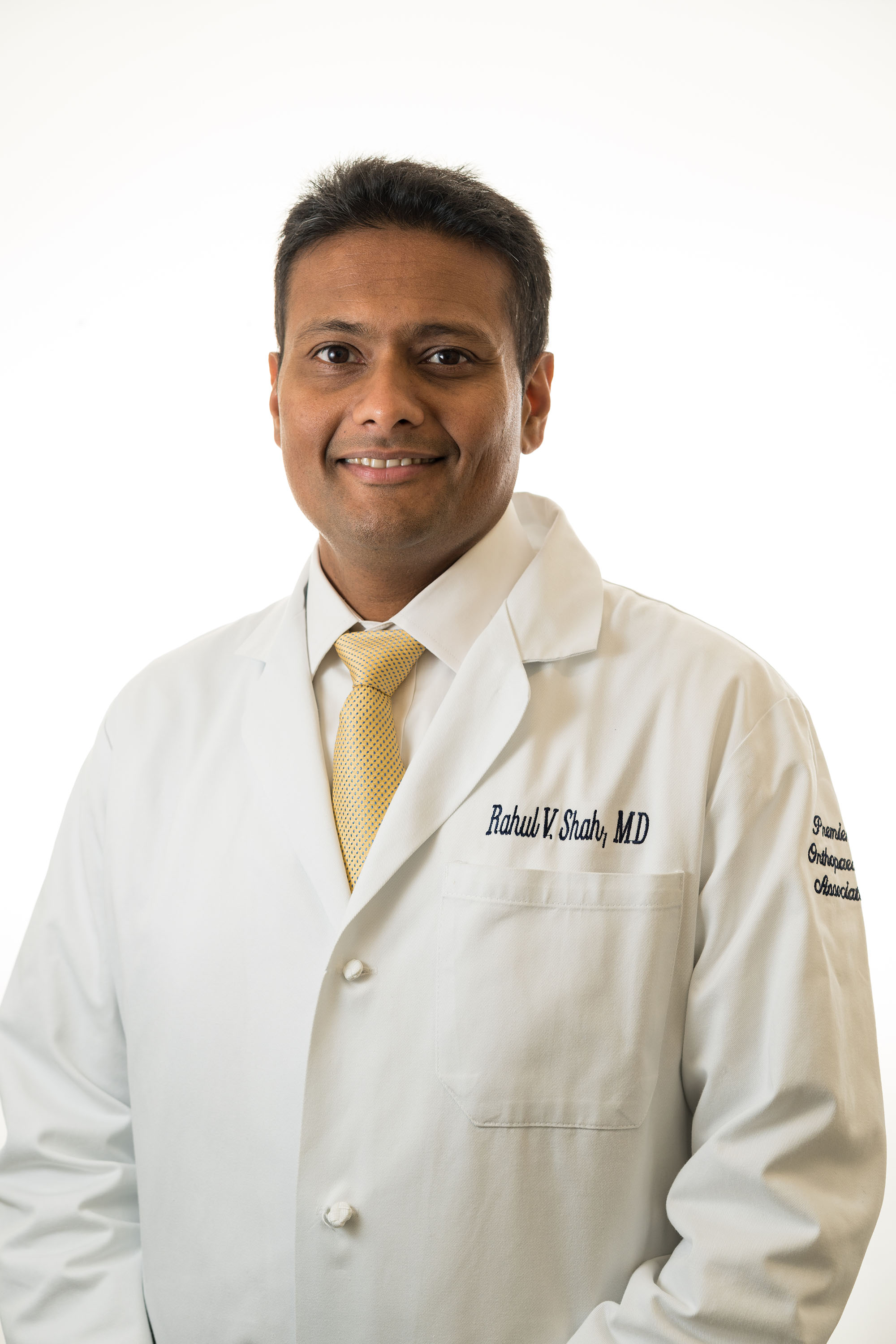
Rahul V. Shah

Dr. Rahul V. Shah, MD, FAAOS

Board-Certified Orthopaedic Spine Surgeon  |  Fellowship-Trained, UCSF  |  Chief Resident, Yale

Dr. Shah performs microdiscectomy with the precision that his UCSF fellowship in minimally invasive spine surgery is built on. He explains to Cherry Hill patients before the procedure exactly what the imaging shows, where the nerve is being compressed, what he will remove, and what the recovery sequence looks like. Patients who go into a microdiscectomy with Dr. Shah understand their own anatomy — which tends to produce better compliance with recovery protocols and better outcomes.

  • FAAOS - Fellow, American Academy of Orthopaedic Surgeons
  • Spine Surgery Fellowship - University of California, San Francisco
  • Chief Resident - Yale University Orthopaedic Surgery
  • Co-Inventor - Stablimax-NZ Dynamic Stabilization Device
  • Faculty - Rowan University Medical School
  • Top Doctor - New Jersey (Castle Connolly)

View Full Profile

Joseph R. Zerbo male in white lab coat

Dr. Joseph R. Zerbo, DO

Board-Certified Orthopaedic Spine Surgeon  |  NASS Member  |  35+ Years in Southern NJ

Dr. Zerbo has performed lumbar microdiscectomy in Camden County for decades and has seen the full range of herniated disc presentations — from the young patient with an acute high-grade herniation to the middle-aged patient with a smaller herniation that has simply not responded to conservative care. His judgment about when the conservative trial has been honestly exhausted and when surgical referral is appropriate is well-calibrated from long experience. Patients at Millennium Surgical Center who receive microdiscectomy from Dr. Zerbo are in familiar hands.

  • AOBOS Board-Certified - American Osteopathic Board of Orthopaedic Surgeons
  • NASS Member - North American Spine Society (since 1999)
  • Chief Resident - Kennedy Memorial Hospital / UMDNJ
  • Founder - Zerbo Spine, PA
  • 35+ Years Serving Southern New Jersey

View Full Profile

Microdiscectomy FAQs

How long will I be out of work after microdiscectomy?

Will my sciatica come back after microdiscectomy?

Is microdiscectomy truly minimally invasive?

Do I need physical therapy after microdiscectomy?

How long will I be out of work after microdiscectomy?

It depends significantly on the type of work. Patients with desk or sedentary jobs typically return to work within two to three weeks. Those with moderate physical demands may return within four to six weeks with activity restrictions. Patients in heavy labor roles — prolonged lifting, prolonged bending, or physically demanding trades — generally need eight to twelve weeks before full return. At our Cherry Hill office, return-to-work planning is part of the pre-operative discussion so patients have realistic expectations.

Will my sciatica come back after microdiscectomy?

The disc fragment removed during microdiscectomy is gone permanently. However, the disc itself remains, and there is a roughly 5–10% lifetime risk of re-herniation at the same level. If re-herniation occurs, the symptoms are typically similar to the original presentation. Revision microdiscectomy is feasible in most cases, though the risk profile is somewhat higher. The majority of patients — approximately 90–95% — do not re-herniate and have durable long-term relief.

Is microdiscectomy truly minimally invasive?

The term 'minimally invasive' is sometimes overused in spine surgery, but microdiscectomy genuinely earns it. The incision is small (typically 1 to 1.5 inches), the approach uses muscle-sparing retraction rather than wide dissection, surgical magnification allows precise work through a limited exposure, and the structural elements of the spine — disc, facet joints, ligaments — are largely preserved. The result is a faster, less painful recovery compared to earlier open disc surgery approaches.

Do I need physical therapy after microdiscectomy?

Yes, and it makes a meaningful difference in outcomes. Physical therapy after microdiscectomy is not about healing the surgical site — the decompression is complete at the time of surgery. It is about rehabilitating the lumbar spine: restoring normal movement patterns, strengthening the core and stabilizing musculature, and reducing the posture and movement habits that may have contributed to the original herniation. Patients who engage consistently with post-operative physical therapy have lower re-herniation rates and better long-term function.

Premier Orthopaedic Associates Cherry Hill

Contact

(856) 690-1616

Hours of Operation

MON — FRI: 8:30AM - 5PM

Contact This Location
Contact us media
Accessibility: If you are vision-impaired or have some other impairment covered by the Americans with Disabilities Act or a similar law, and you wish to discuss potential accommodations related to using this website, please contact our Accessibility Manager at (856) 690-1616.
How can I help you?

Hey there, how can I help you?

Schedule a Visit Find a Provider Bill Pay
Contact Us